Lysosome
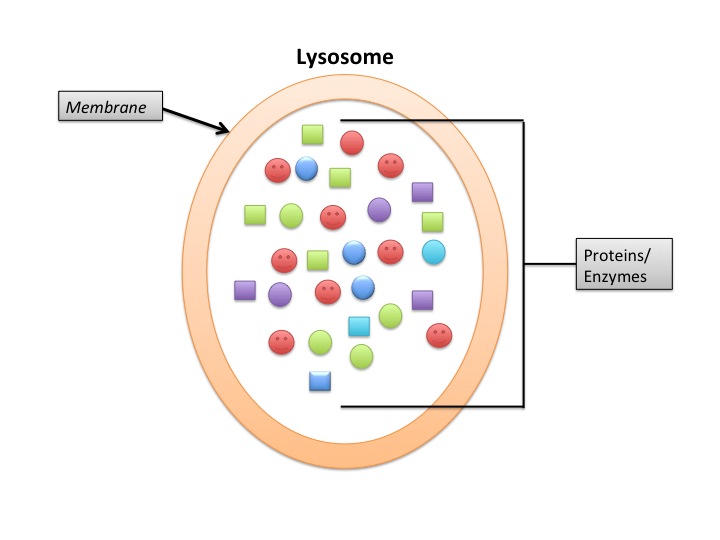
Membrane-bound organelles that serve as the cell's degradation center.

Courtesy of NIAID Ryan Kissinger · Public domain
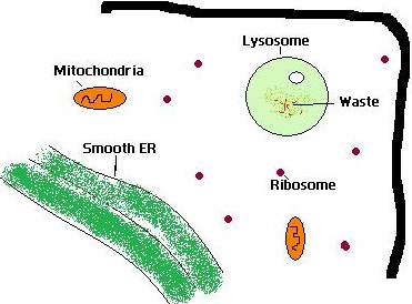
Lysosomes are membrane-bound organelles found in most animal cells, though they are absent from mature mammalian red blood cells; however, some animal red blood cells (e.g., in birds and fish) are nucleated and may contain lysosomes. Plant cells do not have true lysosomes—they rely on vacuoles for similar degradative functions. Lysosomes function as the cell's degradation center, breaking down proteins, polysaccharides, and lipids into their building-block molecules using over 60 different hydrolases in an acidic environment.
- discovered_by
- Christian de Duve
- discovery_period
- 1950s
- location_found
- Laboratory of Physiological Chemistry at the Catholic University of Louvain
- primary_function
- Catabolic degradation of biomolecules
- key_enzymes
- Proteases, glycosidases, lipases, acid phosphatase
- typical_size_range
- 0.1–1.2 μm
- typical_pH_range
- ~4.5–5.0
Lore & Background
Christian de Duve, a Belgian scientist at the Laboratory of Physiological Chemistry at the Catholic University of Louvain, is credited with discovering lysosomes in the 1950s. De Duve and his team were studying the distribution of hydrolytic enzymes such as acid phosphatase within cells, using cell fractionation methods to isolate subcellular components. They identified an unknown organelle rich in acid phosphatase, leading them to propose the existence of lysosomes as membrane-bound organelles containing digestive enzymes. Using differential centrifugation and enzyme activity assays, the team confirmed the hypothesis and understood that these organelles play a crucial role in intracellular digestion processes, such as phagocytosis and autophagy. The presence of digestive enzymes was further validated using electron microscopy.
Reader's Guide
Lysosomes are essential for cellular homeostasis, mediating degradation of both extracellular and intracellular materials through endocytosis and autophagy. They also participate in plasma membrane repair, energy metabolism, cell signaling, and immune response. Their acidic lumen, maintained by proton pumps and chloride antiporters, provides optimal conditions for hydrolases. Lysosomes vary in shape and size, and their numbers can drop below 50 per cell during nutrient deprivation. They sense nutrient availability and regulate mTOR signaling. Lysosomal dysfunction can lead to accumulation of undigested materials, underlying various disorders.
Did You Know?
- Lysosomes are found in most animal cells, but are absent from mature mammalian red blood cells; some animal red blood cells (e.g., in birds and fish) are nucleated and may contain lysosomes.
- The lysosomal lumen has a pH of ~4.5–5.0, maintained by vacuolar-ATPases and ClC-7 antiporters.
- Lysosomes can fuse with the plasma membrane and secrete their contents via lysosomal exocytosis.
- Chaperone-mediated autophagy selectively degrades proteins containing a KFERQ motif.
The Discovery That Redefined Cellular Biology
In the 1950s, a Belgian researcher named Christian de Duve, working at the Laboratory of Physiological Chemistry at the Catholic University of Louvain, set out to map where hydrolytic enzymes like acid phosphatase were distributed inside cells. Using cell fractionation techniques to separate subcellular components, his team stumbled upon something unexpected: an unidentified organelle packed with acid phosphatase. Rather than dismissing it as a curiosity, de Duve and colleagues proposed that this structure was a dedicated, membrane-enclosed compartment housing digestive enzymes capable of breaking down a wide range of biological molecules. They confirmed their hypothesis through differential centrifugation, enzyme activity assays, and ultimately electron microscopy, which revealed the organelle's physical form. Their work illuminated the lysosome's central role in intracellular digestion, including phagocytosis and autophagy. The discovery opened an entirely new field of inquiry into lysosomal function and the devastating disorders that arise when undigested material accumulates within cells. In 1974, de Duve was honored with the Nobel Prize in Physiology or Medicine for this landmark contribution.
An Acid Fortress at the Heart of the Cell
Found in virtually every animal cell (with the notable exception of red blood cells) and only rarely in plant cells, the lysosome is a small, membrane-bound compartment that typically numbers in the hundreds within a single cytosol. Its shape is anything but uniform: depending on the cell type and what it is currently digesting, a lysosome may appear spherical, ovoid, or even tubular, with diameters ranging from 0.1 to 1.2 micrometers and tubular variants stretching as far as 15 micrometers in phagocytes. The defining feature of this organelle is its interior chemistry. A single-bilayer phospholipid membrane, studded with heavily glycosylated proteins that form a protective glycocalyx, encloses a lumen whose pH hovers around 4.5 to 5.0—sharply acidic compared to the slightly basic cytosol at pH 7.2. This acidity is the precise condition under which the more than sixty hydrolases inside operate at peak efficiency. Because these enzymes are pH-sensitive and essentially inert in the alkaline cytosol, the membrane acts as a critical safety barrier, preventing the cell from digesting itself should any hydrolases leak out. To sustain the acidic gradient, vacuolar-ATPase proton pumps shuttle hydrogen ions from mitochondria into the lumen, while the ClC-7 antiporter counter-transports chloride ions, maintaining both the pH and ionic balance essential for the lysosome's work.
Two Roads Into the Digestive Machine
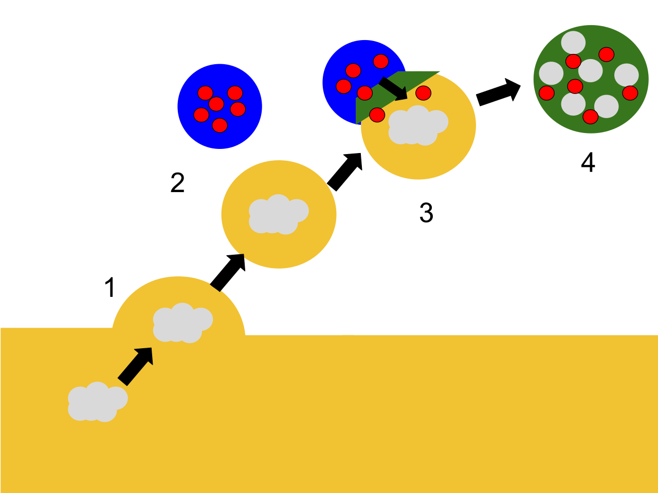
The lysosome does not simply sit idle waiting for cargo; it actively cycles through fusion and fission events with partner organelles, serving as a reservoir of acidic hydrolases. Material arrives along two principal routes. The first is the endocytic pathway, through which extracellular particles—complex lipids, membrane proteins, polysaccharides—travel from early endosomes into late endosomes, also called multivesicular bodies. From there, the late endosome meets the lysosome either through full membrane fusion or through a subtler kiss-and-run interaction, in which the two organelles briefly touch, exchange contents, and separate again. The resulting hybrid, an endolysosome, is where the actual breakdown of endocytic cargo takes place under normal conditions. The second route is autophagy. Damaged organelles and misfolded proteins are sequestered into autophagosomes, which then merge with lysosomes to form autolysosomes. Inside these structures, acidic hydrolases dismantle the cargo into amino acids, monosaccharides, and free fatty acids. Once degradation is complete, the resulting building blocks are shuttled back into the cytosol through specific membrane transport proteins or vesicular trafficking, ready to be recycled into new cellular structures or burned for energy. Remarkably, lysosomes can also fuse directly with the plasma membrane in a process called lysosomal exocytosis, releasing their contents outside the cell.
Guardian, Sentinel, and Metabolic Sensor
Beyond its well-known role as a degradation center, the lysosome is deeply woven into the cell's defense and regulatory networks. In macrophages, lysosomes team up with phagosomes to kill and digest invading bacteria and virus particles, effectively serving as the immune system's intracellular killing chamber. They also house toll-like receptors such as TLR7 and TLR9, which help detect pathogens and trigger downstream signaling. Degraded microbial material can be processed into antigen fragments, loaded onto MHC molecules, and presented to T-cells—a critical step in immune defense. The lysosome's influence extends to cell fate itself: if its hydrolytic enzymes are released into the cytoplasm, they can trigger a form of programmed cell death known as lysosomal-mediated programmed cell death, or LM-PCD. On the metabolic front, lysosomes act as nutrient sensors. When the cell is well-fed, they activate mTOR signaling to promote anabolic, biosynthetic processes. During starvation, they shift gears, degrading autophagic material and recycling the resulting components to keep the cell alive. They also contribute to plasma membrane repair, cell homeostasis, energy metabolism, and cell signaling, making them far more than a simple waste-disposal unit.
Gallery
Frequently Asked Questions
Who is Lysosome?
Lysosome was first identified in the 1950s by biochemist Christian de Duve, working out of the Laboratory of Physiological Chemistry at the Catholic University of Louvain. It is a membrane-enclosed organelle present in the vast majority of animal cells.
What are Lysosome's powers/role?
Lysosome serves as the cell's dedicated recycling and waste-disposal station, digesting proteins, complex carbohydrates, and fats back into their constituent monomers. It carries out this work through more than sixty distinct hydrolase enzymes operating in a highly acidic interior.
Where does Lysosome appear in the story?
You will find Lysosome in most animal cell types, though it is notably absent from mature mammalian red blood cells; nucleated red blood cells in birds and fish, however, can still house them. Plant cells skip Lysosome entirely, relying on their large central vacuole to handle comparable breakdown tasks.
How big is Lysosome?
Individual Lysosome units typically range from 0.1 to 1.2 micrometers in diameter, making them one of the smaller membrane-bound organelles you will encounter inside a cell.
Why is Lysosome important?
Without Lysosome, cells would accumulate undigested macromolecules and lose the ability to recycle amino acids, sugars, and fatty acids for new biosynthetic pathways. Its arsenal of proteases, glycosidases, lipases, and acid phosphatase makes it indispensable for maintaining cellular homeostasis and clearing out damaged organelles.
More in Cellular And Molecular Biology 1-24
Elsewhere in the Cellular And Molecular Biology universe
Spotted an error? Know more?
This is a living reference — every entry is fact-audited, and reader corrections feed straight into our audit queue. Suggest an edit · See this site's audit record