
Membrane potential
The electric potential difference across a biological cell membrane.

Д.Ильин : vectorization · CC0
Membrane potential, also known as transmembrane potential or membrane voltage, is the difference in electric potential between the interior and the exterior of a biological cell. By convention, it is written as Vm = Vinside − Voutside, so a negative membrane potential means the cell interior is negative relative to the outside. This potential is the energy per charge required to move a very small positive charge at constant velocity across the cell membrane from the exterior to the interior.
- field
- Cell biology, electrophysiology
- known_for
- Difference in electric potential across a cell membrane; basis for cellular signaling and battery-like function
- resting_potential_neurons
- −80 to −70 mV
- units
- millivolts (mV)
Lore & Background
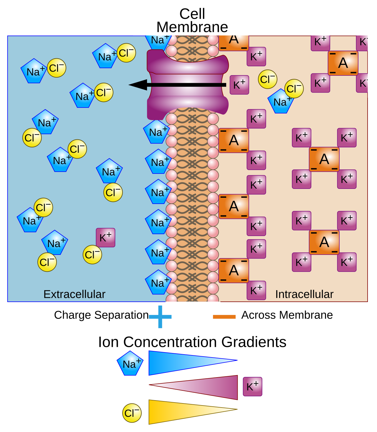
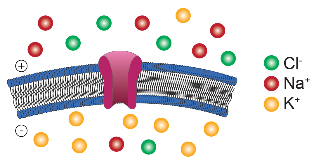
All animal cells are surrounded by a membrane composed of a lipid bilayer with embedded proteins. This membrane serves both as a capacitor and as a diffusion barrier to ion movement. Ion pumps actively establish concentration gradients, and ion channels allow ions to move down their electrochemical gradients. These transmembrane proteins are electrically analogous to resistors and current sources, contributing to the voltage across the membrane. The membrane potential allows a cell to function as a battery, powering molecular devices, and in excitable cells like neurons and muscle cells, it is used for transmitting signals. Signals in excitable cells are generated by opening or closing ion channels at one point, producing a local change in membrane potential. This change can be sensed by adjacent or distant ion channels, which then open or close, reproducing the signal. In baseline states, the membrane potential is held at a stable resting potential—for neurons, ranging from −80 to −70 mV. Depolarization occurs when the interior voltage becomes less negative, and hyperpolarization when it becomes more negative. A sufficiently large depolarization can evoke an action potential, in which the membrane potential changes rapidly and significantly, often reversing polarity. Differences in ion concentrations across the membrane lead to the membrane potential. Potassium (K+) is high inside and low outside; sodium (Na+) and chloride (Cl−) are high outside and low inside. If the membrane is selectively permeable to potassium, these ions diffuse down their gradient, leaving uncompensated negative charges inside. This separation of charges, which lines up on the membrane surfaces, is the basis of the membrane voltage. The system as a whole is electro-neutral, and the voltage is physically located only in the immediate vicinity of the membrane.
Reader's Guide
Membrane potential is a fundamental concept in cell biology, describing the voltage difference across a cell's plasma membrane. Its significance lies in two basic functions: it allows a cell to act as a battery, powering molecular devices embedded in the membrane, and it enables signal transmission in excitable cells such as neurons and muscle cells. The potential arises from concentration gradients of ions like potassium, sodium, and chloride, combined with selective membrane permeability. The physical basis involves electrical force and diffusion. In neurons, the resting potential is typically −80 to −70 mV, and changes in this potential—depolarization or hyperpolarization—underlie electrical signaling. Action potentials, rapid and significant voltage changes, are generated by voltage-gated ion channels and can reverse polarity. The membrane potential is measured by convention with the outside set to zero, and the interior potential relative to that. Its legacy is central to understanding nerve impulses, muscle contraction, and cellular homeostasis, and it remains a key topic in physiology and neuroscience.
Did You Know?
- By convention, membrane potential is defined as Vm = Vinside − Voutside, so a negative value means the interior is negative relative to the exterior.
- In neurons, the resting potential is defined as ranging from −80 to −70 millivolts.
Defining the Voltage Across a Living Boundary
The membrane potential represents the electric potential difference between a cell's interior and its external environment. By standard convention, this value is expressed as the interior potential minus the exterior potential, meaning a negative reading tells us the inside sits at a lower voltage than the outside. In practical terms, this potential quantifies the energy per unit charge needed to shuttle a tiny positive test charge at steady speed from the exterior to the interior through the membrane. Of course, if that charge were permitted to accelerate, additional considerations such as kinetic energy shifts and electromagnetic radiation would enter the picture. Typical readings across animal cells land in the tens-of-millivolts range, with values commonly spanning roughly minus twenty to minus two hundred millivolts depending on the particular cell type and its physiological state. For these typical negative values, pushing a positive charge from inside to outside demands positive work, yet the random thermal motion of ions provides enough kinetic energy for some to surmount the barrier, enabling a net ionic flow even against the gradient when the membrane is selectively permeable.
The Membrane as a Molecular Capacitor and Battery
Every animal cell is encased in a lipid bilayer studded with transmembrane proteins, and this structure simultaneously acts as a capacitor and as a selective diffusion barrier for ions. Ion pumps embedded in the bilayer actively transport ions against their gradients, establishing the concentration differences that underpin the voltage, while ion channels permit ions to drift down their electrochemical gradients. Electrically, the channels behave like resistors and the pumps like current sources, together generating the voltage that spans the two faces of the membrane. Because the bilayer is only a few nanometers thick, even a modest potential of one hundred millivolts produces an extraordinarily intense electric field right at the membrane surface. The membrane potential serves two fundamental roles: it lets the cell operate as a biological battery, powering a variety of molecular devices embedded in the lipid sheet, and in electrically excitable cells such as neurons and muscle fibers, it provides the mechanism for transmitting electrical signals from one region of the cell to another.
From Resting State to Action Potential
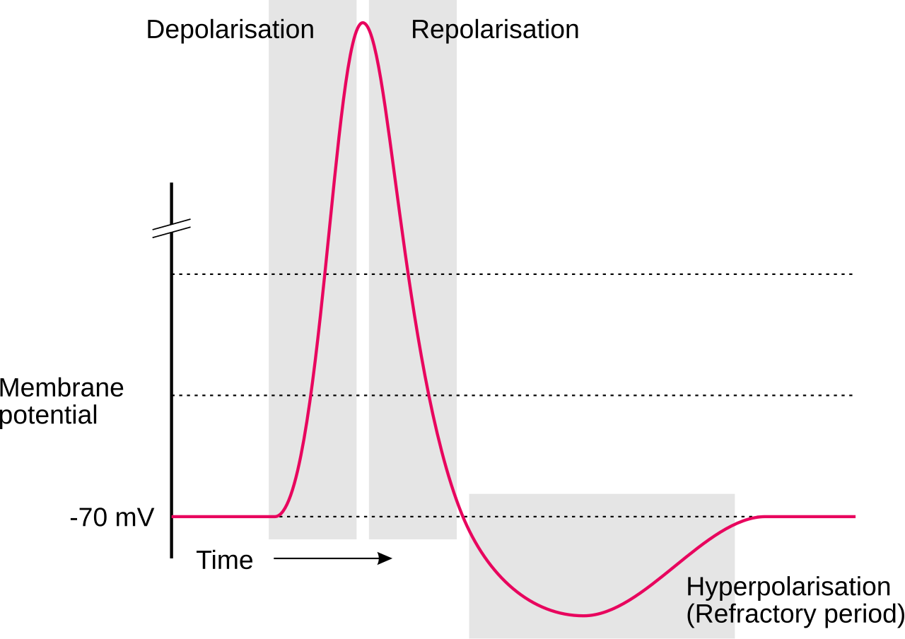
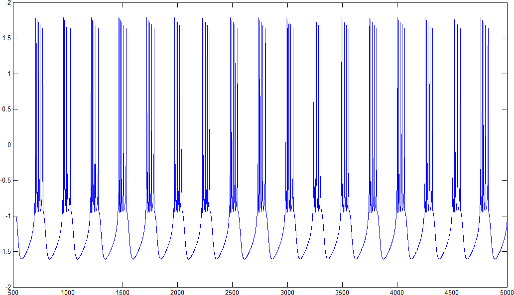
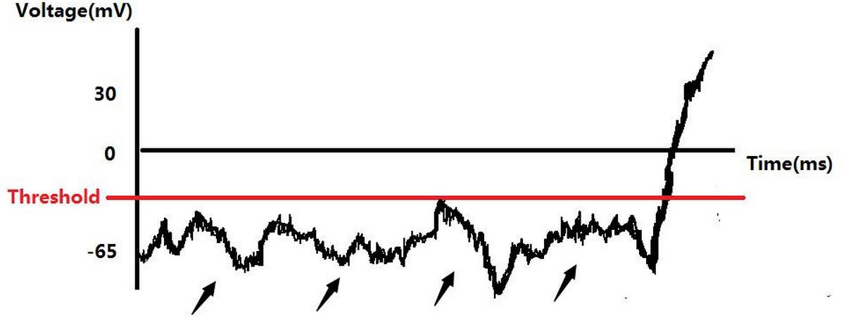
In neurons, the baseline membrane potential, known as the resting potential, typically hovers between minus eighty and minus seventy millivolts, meaning the interior carries a negative charge of a bit under one-tenth of a volt relative to the outside. When ion channels open or close at a particular spot on the membrane, the local potential shifts. If the interior voltage becomes less negative, the event is termed a depolarization, such as moving from minus seventy to minus sixty millivolts; if it becomes more negative, the shift is called a hyperpolarization, like going from minus seventy to minus eighty. In excitable cells, a depolarization large enough can trigger an action potential, a rapid and dramatic reversal of polarity lasting anywhere from one to one hundred milliseconds. These events are driven by voltage-gated ion channels. Because those channels are themselves regulated by the membrane potential, which in turn is shaped by the channels, feedback loops emerge that produce rich temporal dynamics, including oscillations and regenerative cascades.
Charge Separation and the Geometry of the Voltage
Although the entire cell interior is described as negative, the bulk fluids on both sides of the membrane remain electrically neutral. The voltage exists solely because of a thin sheet of charge that accumulates at the membrane surfaces. In the simplest illustrative case, if the membrane is selectively permeable to potassium, those positively charged ions diffuse outward down their concentration gradient, leaving behind uncompensated negative charges inside. The uncompensated positive charges outside and the negative charges inside physically align on opposite faces of the bilayer and attract one another across the lipid layer. Thus the membrane potential is not a property of the whole cell volume but is physically confined to the immediate vicinity of the membrane. Potassium dominates this picture because of its high permeability, while sodium, chloride, calcium, and other ions, despite possessing strong concentration gradients, contribute more modestly because the membrane is less permeable to them.
Gallery
Frequently Asked Questions
Who is Membrane potential?
Membrane potential (Vm) is the voltage gap between a cell's cytoplasm and its extracellular surroundings, defined as the interior potential minus the exterior. It is reported in millivolts, and a negative value simply means the inside of the cell sits at a lower electric potential than the fluid outside.
What are Membrane potential's powers and role?
It functions as a built-in cellular battery, storing electrochemical energy that powers nerve impulses, muscle contractions, and selective ion transport. Without this gradient, cells would lack the driving force needed for electrical signaling and osmotic balance.
How does Membrane potential's story end?
In a living cell the narrative is ongoing: Na⁺/K⁺-ATPase pumps and leak channels constantly restore the gradient after each signaling event. In a dying cell the pumps shut down, the gradient dissipates toward zero, and electrical excitability is lost for good.
What units and conventions does Membrane potential use?
It is always expressed in millivolts (mV) and written with the convention Vm = Vinside − Voutside. A negative reading is not an error; it is the expected sign indicating the cytoplasm is more electronegative than the bath outside the membrane.
More in Cellular And Molecular Biology 1-24
Elsewhere in the Cellular And Molecular Biology universe
Spotted an error? Know more?
This is a living reference — every entry is fact-audited, and reader corrections feed straight into our audit queue. Suggest an edit · See this site's audit record