
Lipid bilayer
Thin polar membrane forming a barrier around all cells.

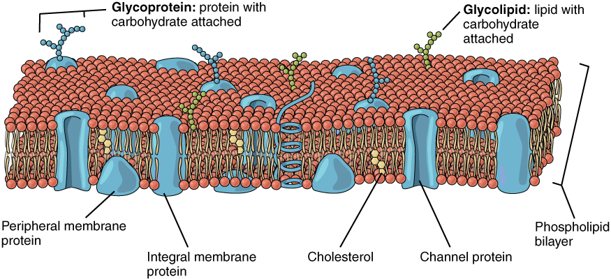
Original: Stephen Gilbert Vector: derivative work: Urutseg · CC BY-SA 3.0
The lipid bilayer (or phospholipid bilayer) is a thin polar membrane made of two layers of lipid molecules. These membranes form a continuous barrier around all cells, and are found in the cell membranes of almost all organisms and many viruses, as well as in the nuclear membrane and membranes of membrane-bound organelles. The bilayer is the barrier that keeps ions, proteins, and other molecules where they are needed and prevents them from diffusing into areas where they should not be, making it essential for cellular compartmentalization and regulation.
- field
- Cell biology, biophysics
- known_for
- Forming the fundamental barrier of all cell membranes
- composition
- Two layers of amphiphilic phospholipids with hydrophilic heads and hydrophobic tails
- thickness
- Hydrophobic core typically 3-4 nm thick; total bilayer a few nanometers
- key_property
- Impermeable to most water-soluble molecules and ions
Lore & Background
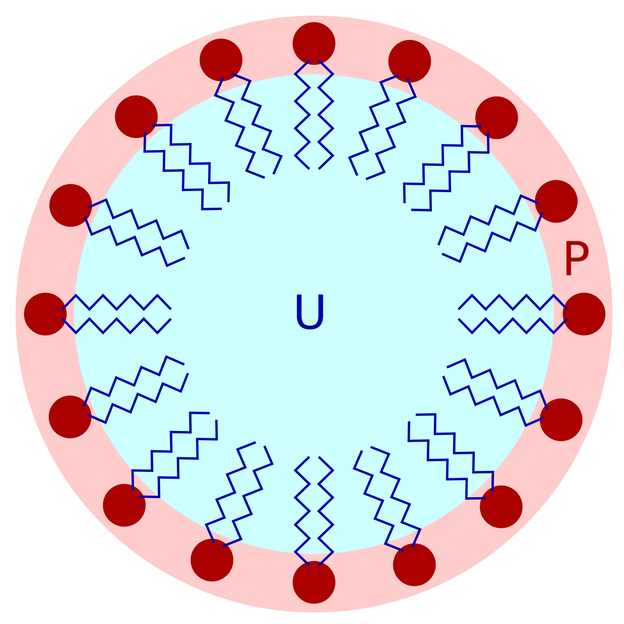
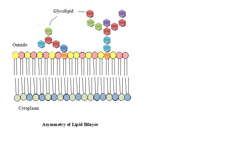
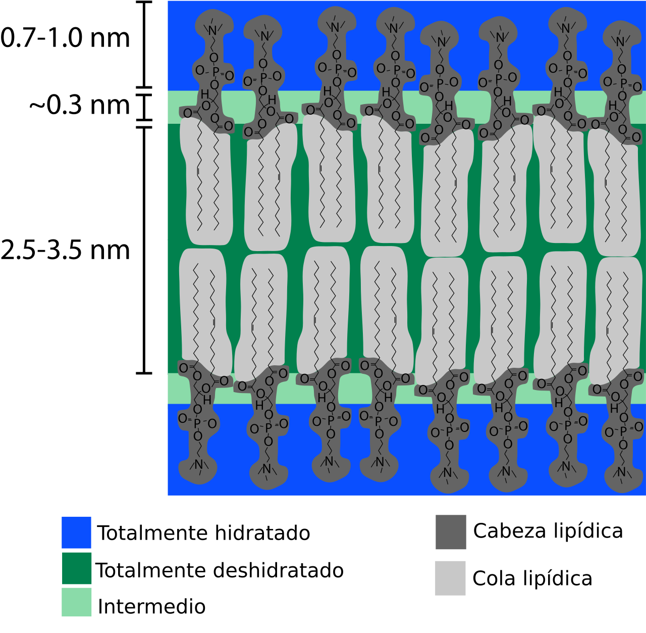
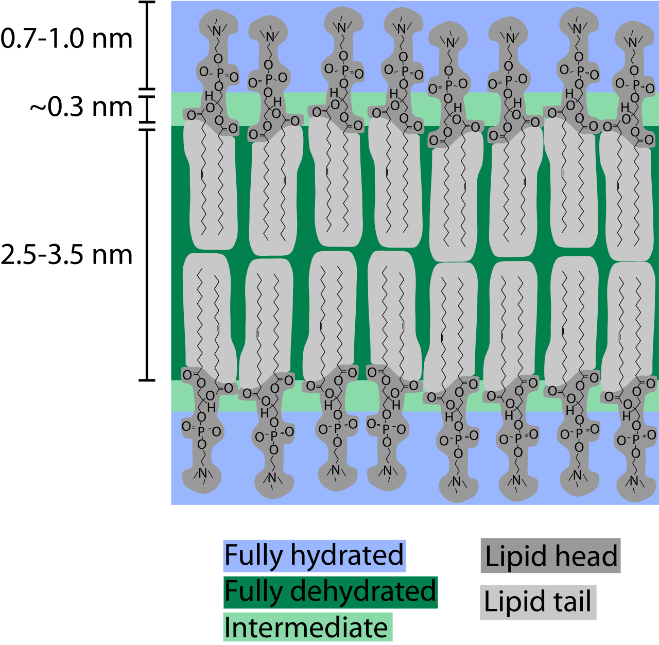
When phospholipids are exposed to water, they self-assemble into a two-layered sheet with hydrophobic tails pointing toward the center, creating two leaflets. This assembly is driven by the hydrophobic effect and includes non-covalent interactions such as van der Waals forces, electrostatic and hydrogen bonds. The bilayer is very thin compared to its lateral dimensions; if a typical mammalian cell were magnified to the size of a watermelon, the plasma membrane would be about as thick as a piece of office paper. The bilayer's cross-section includes a hydrophilic headgroup region (0.8-0.9 nm thick), a partially hydrated boundary layer (about 0.3 nm thick), and a hydrophobic core (3-4 nm thick). In many naturally occurring bilayers, the compositions of the inner and outer leaflets are different. For example, in human red blood cells, the inner leaflet is composed mostly of phosphatidylethanolamine, phosphatidylserine, and phosphatidylinositol, while the outer leaflet is based on phosphatidylcholine, sphingomyelin, and glycolipids. This asymmetry arises partly because most phospholipids are synthesized and initially inserted into the inner monolayer, with some transported to the outer monolayer by enzymes called flippases. Other lipids, such as sphingomyelin, are synthesized in the Golgi apparatus and then transported to the outer leaflet. Lipid asymmetry does not normally dissipate quickly because spontaneous flip-flop of lipids between leaflets is extremely slow. At a given temperature, a lipid bilayer can exist in either a liquid or a gel (solid) phase. All lipids have a characteristic temperature at which they transition from gel to liquid phase. In liquid phase bilayers, lipids exchange locations with neighbors millions of times a second, allowing diffusion across the membrane surface, whereas in gel phase bilayers, lipids have less mobility. The bilayer can adopt a solid gel phase at lower temperatures and undergo phase transition to a fluid state at higher temperatures, with the chemical properties of the lipid tails influencing the transition temperature.
Reader's Guide
The lipid bilayer is of fundamental significance because it defines the boundary of all cells and their internal compartments, enabling life at the cellular level. Its impermeability to ions and most water-soluble molecules allows cells to regulate salt concentrations and pH by transporting ions across membranes using ion pumps. The bilayer's structure, composed of amphiphilic phospholipids, provides a versatile platform: phospholipid head groups can alter surface chemistry and serve as signals or anchors for other molecules, while lipid tails affect membrane properties such as phase behavior and mechanical resistance to stretching and bending. Cholesterol, present in animal cells, helps strengthen the bilayer and decrease its permeability, and also helps regulate the activity of certain integral membrane proteins. These membrane proteins, held tightly by an annular lipid shell, are involved in many intra- and inter-cellular signaling processes, including membrane fusion events such as the acrosome reaction during fertilization or viral entry into a cell. The study of bilayers has advanced through techniques like x-ray reflectometry, neutron scattering, nuclear magnetic resonance, electron microscopy, and atomic force microscopy, as well as through artificial model bilayers used in drug delivery vesicles. The asymmetry of natural bilayers, maintained by enzymes like flippases and scramblases, plays roles in processes such as apoptosis, where phosphatidylserine exposure signals macrophages to scavenge dying cells.
Did You Know?
- The lipid bilayer is only a few nanometers thick, yet it is impermeable to most water-soluble molecules and ions.
- In human red blood cells, the inner and outer leaflets of the bilayer have different lipid compositions.
- Cholesterol helps strengthen the bilayer and decrease its permeability in animal cells.
- Lipid bilayers can adopt a solid gel phase at lower temperatures and a fluid phase at higher temperatures.
Self-Assembly and Cross-Sectional Architecture
When phospholipids encounter an aqueous environment, their dual nature drives them into a remarkably ordered arrangement. Each molecule carries a water-loving phosphate head alongside two water-averse fatty acid chains. In water, these molecules spontaneously organize into a two-leaflet sheet, with all hydrophobic tails tucked toward the interior and heads facing the surrounding solution. This self-assembly is powered by the hydrophobic effect and reinforced by non-covalent forces including van der Waals interactions, electrostatic attractions, and hydrogen bonding. The resulting structure is astonishingly thin relative to its lateral spread. If a typical mammalian cell were scaled up to the size of a watermelon, its plasma membrane would be roughly as thick as a sheet of office paper. Yet even within those few nanometers, distinct chemical zones exist. The fully hydrated headgroup region spans about 0.8 to 0.9 nanometers, followed by a partially hydrated boundary layer of roughly 0.3 nanometers where water concentration plummets from 2 molar to nearly zero. Beyond that lies the hydrophobic core, typically 3 to 4 nanometers thick, whose exact dimensions shift with chain length, chemical composition, and temperature near phase transitions.
Selective Permeability and Ion Homeostasis
The lipid bilayer serves as the fundamental boundary for virtually every cell on Earth, as well as for many viruses, the nuclear envelope, and the membranes of internal organelles. Despite being only a few nanometers wide, this barrier is extraordinarily effective at blocking the passage of water-soluble molecules. The hydrophobic interior of the bilayer presents a formidable obstacle to ions, sugars, and other hydrophilic solutes that cannot simply dissolve through the fatty acid core. This selective impermeability is what gives cells the ability to maintain distinct internal environments. By controlling which ions cross the membrane, cells can regulate their salt concentrations and pH levels. Specialized proteins known as ion pumps handle the active transport of charged particles across this otherwise impenetrable wall. In animal cells, cholesterol plays a critical supporting role. Embedded among the phospholipids, it reinforces the bilayer's structural integrity, further reduces its permeability, and fine-tunes the activity of integral membrane proteins that are anchored within the lipid environment by an annular shell of surrounding lipids. Together, these features make the bilayer not merely a passive wall but a dynamically regulated gateway.
Asymmetry, Signaling, and Membrane Fusion
In many living cells, the two leaflets of the bilayer are not mirror images of one another. Human red blood cells illustrate this clearly: the cytoplasmic leaflet is dominated by phosphatidylethanolamine, phosphatidylserine, and phosphatidylinositol derivatives, while the extracellular leaflet is built primarily from phosphatidylcholine, sphingomyelin, and assorted glycolipids. This compositional asymmetry partly reflects where each lipid is originally synthesized within the cell. Its biological significance is still being unraveled, but one well-established role involves programmed cell death. During apoptosis, phosphatidylserine migrates from the inner leaflet to the outer surface, where it is detected by macrophages that then engulf and clear the dying cell. Beyond asymmetry, the chemical identity of lipid head groups can serve as signaling cues and molecular anchors for other membrane components, while the fatty acid tails govern whether the bilayer exists in a solid gel or fluid phase. Certain specialized membrane proteins mediate the fusion of two separate bilayers, a process essential for events as diverse as the acrosome reaction during fertilization and the entry of a virus into a host cell.
Experimental Challenges and Clinical Applications
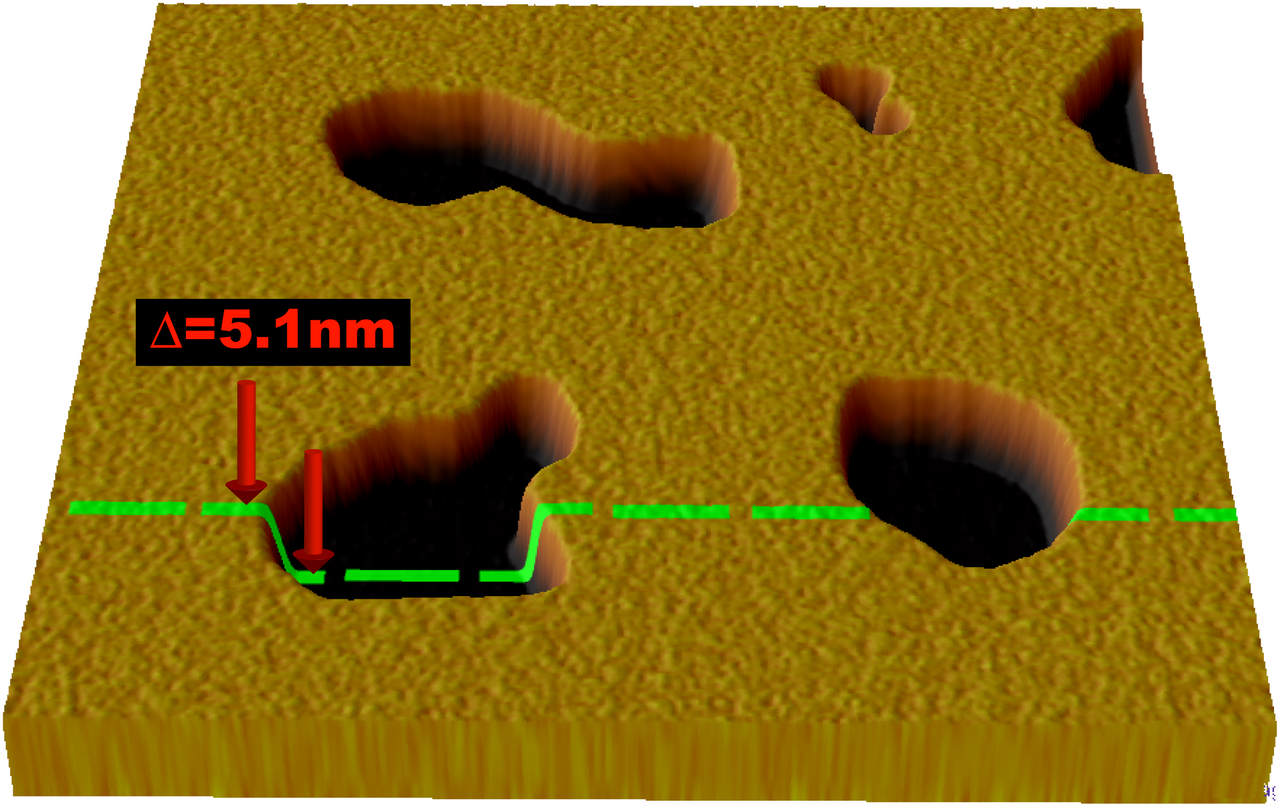
Studying the lipid bilayer presents unique difficulties. Because these membranes are both extremely fragile and invisible under a traditional microscope, researchers must turn to advanced imaging tools such as electron microscopy and atomic force microscopy to observe them directly. Characterizing the detailed cross-sectional architecture—mapping the hydrated headgroup zone, the partial-hydration boundary, and the hydrophobic core—has relied on sophisticated biophysical techniques including x-ray reflectometry, neutron scattering, and nuclear magnetic resonance spectroscopy. In the laboratory, scientists construct artificial model bilayers to probe fundamental properties like mechanical resistance to stretching and bending, as well as the temperature-dependent phase transitions between solid gel and fluid states. These model systems are not merely academic curiosities. Vesicles formed from such bilayers have found genuine clinical use as drug-delivery vehicles, exploiting the bilayer's natural ability to shield its interior from the external aqueous environment. The packing density of lipids within these artificial membranes also influences their mechanical behavior, making them useful platforms for understanding how real cell membranes resist deformation.
Gallery
Frequently Asked Questions
Who is Lipid bilayer?
The lipid bilayer is the two-layered polar membrane that wraps around every cell and lines the internal compartments of eukaryotic cells. It is the boundary structure that defines where a cell begins and ends, found in nearly all organisms and even many viruses.
What are Lipid bilayer's powers/role?
Its signature ability is acting as a selective barrier: it blocks most water-soluble ions and polar molecules from freely diffusing across, so cells must rely on dedicated transport proteins to move substances in and out. This is what makes cellular compartmentalization and metabolic regulation possible.
How does Lipid bilayer's story end?
There is no single dramatic finale; when a cell dies or is lysed, the phospholipid molecules that built the bilayer are enzymatically broken down and their fatty-acid, glycerol, and phosphate components are simply recycled into the organism's general metabolic pool. The organized two-sheet structure simply ceases to exist once the cell is no longer maintained.
Why is Lipid bilayer important?
Without it, cells could not hold distinct internal environments, and the concentration gradients that power energy production, signaling, and transport would collapse. It is the single most universal structural feature shared by virtually all known cells, making it foundational to life as we understand it.
What is Lipid bilayer made of and how thick is it?
It is built from two parallel sheets of amphiphilic phospholipids, with hydrophilic head groups facing the watery environments on either side and hydrophobic fatty-acid tails tucked into the interior. The hydrophobic core spans roughly 3–4 nanometers, so the entire bilayer is only a few nanometers thick.
More in Cellular And Molecular Biology 1-24
Elsewhere in the Cellular And Molecular Biology universe
Spotted an error? Know more?
This is a living reference — every entry is fact-audited, and reader corrections feed straight into our audit queue. Suggest an edit · See this site's audit record