RNA splicing
Process removing introns to form mature mRNA for translation.

RNA splicing is a molecular biology process in which a precursor messenger RNA (pre-mRNA) transcript is transformed into mature messenger RNA (mRNA) by removing introns (non-coding regions) and joining exons (coding regions). For nuclear-encoded genes, splicing occurs in the nucleus during or immediately after transcription, and is essential for creating mRNA that can be translated into protein. Splicing is a key step in gene expression, the central dogma of molecular biology.
- field
- Molecular biology
- known_for
- Removal of introns and joining of exons in pre-mRNA to produce mature mRNA
- process_type
- Post-transcriptional modification
- catalyzed_by
- Spliceosome (complex of snRNPs) or self-splicing ribozymes
- splice_site_consensus
- GU at 5' end, AG at 3' end of intron
Lore & Background
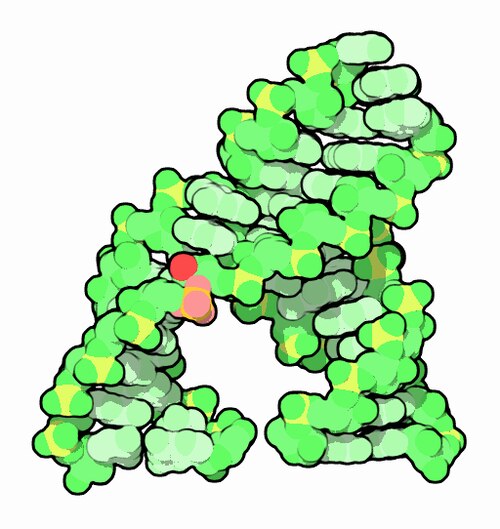
RNA splicing occurs via several pathways, depending on intron structure and catalysts. The spliceosomal complex, composed of five small nuclear ribonucleoproteins (snRNPs), catalyzes splicing for most eukaryotic introns. Assembly and activity of the spliceosome occur during transcription of pre-mRNA. The major spliceosome splices introns with GU-AG boundaries, while the minor spliceosome handles rare introns with AU-AC consensus sequences. Recursive splicing removes very long introns in steps, first found in the Drosophila Ultrabithorax gene. Trans-splicing joins exons from different RNA transcripts.
Reader's Guide
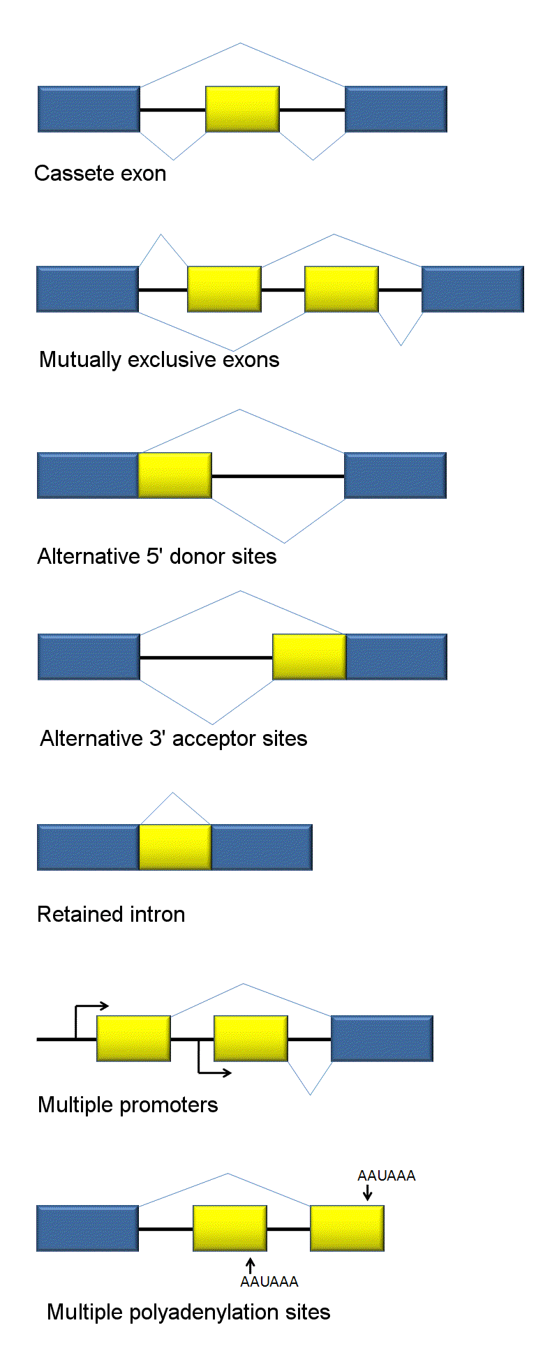
RNA splicing is a fundamental mechanism in gene expression, enabling the production of diverse proteins from a single gene through alternative splicing. The spliceosome's catalytic core resembles self-splicing group II introns, suggesting an evolutionary link to an ancient RNA world. Splicing errors, such as point mutations activating cryptic splice sites, can lead to protein truncations or deletions. Understanding splicing has implications for genetic disorders and therapeutic targeting. The process accounts for over 99% of splicing via the canonical lariat pathway, with noncanonical splicing occurring for rare introns.
Did You Know?
- Self-splicing introns are ribozymes that catalyze their own excision without proteins.
- The major spliceosome uses U1, U2, U4, U5, and U6 snRNPs to splice introns with GU-AG boundaries.
- Recursive splicing removes very long introns in steps, first observed in the Drosophila Ultrabithorax gene.
- Trans-splicing joins exons from two different RNA transcripts, including endogenous and exogenous RNAs.
The Essential Role in Gene Expression
RNA splicing stands as one of the most critical steps between a gene's raw output and a functional protein. When a eukaryotic cell transcribes a nuclear-encoded gene, the initial product is a precursor messenger RNA that still contains non-coding segments called introns interspersed among the coding exons. Unless those introns are excised and the exons are rejoined, the transcript cannot serve as a template for protein synthesis. This processing typically takes place inside the nucleus, either while transcription is still underway or immediately afterward. The entire pipeline—transcription, splicing, and translation—is what molecular biologists refer to as gene expression, the so-called central dogma. While the vast majority of eukaryotic introns rely on a protein-RNA machine called the spliceosome to carry out their removal, a fascinating minority are self-splicing ribozymes, RNA molecules that catalyze their own excision without any protein assistance. In either case, the end goal is the same: a clean, translatable mRNA ready for the ribosome.
The Spliceosome: A Stepwise Molecular Assembly Line
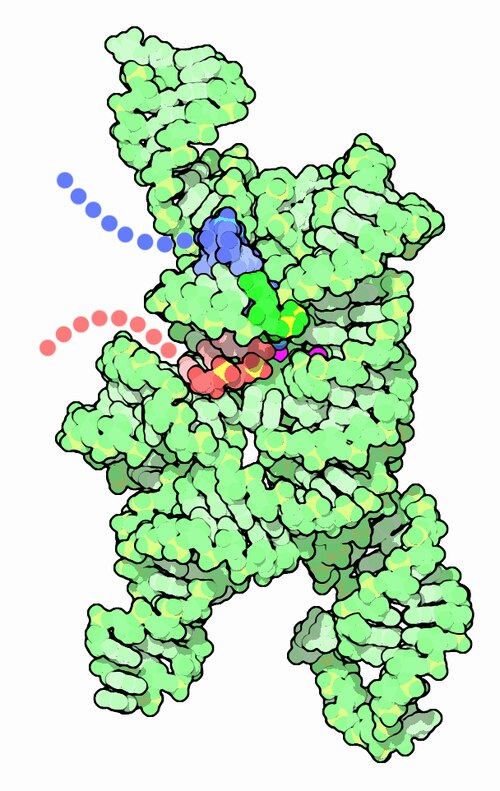
The spliceosome is a massive RNA-protein complex built from five small nuclear ribonucleoproteins, and its assembly unfolds in a carefully choreographed sequence of intermediates. It all begins with Complex E, where the U1 snRNP latches onto the GU dinucleotide at the intron's 5' donor site, SF1 recognizes the branch point, and the U2AF proteins anchor at the 3' acceptor region and the polypyrimidine tract. In Complex A, U2 displaces SF1 at the branch point, consuming ATP. The U4/U5/U6 trimer then docks to form Complex B, with U5 making contact at the 5' exon and U6 pairing with U2. Transitioning to B*, U1 is shed and U6 repositions to the 5' splice site. The catalytic moment arrives in Complex C: U4 is released, and the U6/U2 pair drives transesterification, ligating the 5' intron end to the branch adenine to create the characteristic lariat while cleaving the 5' junction. Finally, in C*, the 3' site is cut, the two exons are ligated using another round of ATP hydrolysis, the spliced RNA and lariat are released, and the snRNPs are recycled. This canonical lariat pathway accounts for over 99% of all splicing events.
Reading the Intron: Splice Site Architecture and the Cost of Errors
The term intron is a contraction of intragenic region and intracistron, reflecting its position between two exons within a gene. These non-coding segments are remarkably widespread, appearing in the genes of most organisms and many viruses, and they are not limited to protein-coding genes—they also inhabit rRNA and tRNA genes. Each spliceosomal intron carries a precise set of recognition landmarks: a nearly invariant GU at the 5' donor site, a polypyrimidine tract rich in cytosines and uridines upstream of the 3' end, an almost invariant AG at the acceptor site, and a branchpoint adenine roughly 20 to 50 nucleotides upstream of that acceptor. The exact spacing between the branchpoint and the acceptor, along with the specific nucleotide composition of these elements, influences which splice site the machinery selects. This sensitivity has real biological consequences. A single point mutation in the underlying DNA, or a transcription error, can activate a cryptic splice site that is normally silent. The result is a mature mRNA missing a chunk of exon sequence, turning what should have been a single amino-acid substitution into a full deletion or truncation of the final protein.
Beyond the Canonical Pathway: Recursive, Trans, and Minor Splicing
Not every intron follows the straightforward GU-AG lariat route. When the flanking sequences deviate from that canonical rule, a distinct minor spliceosome takes over, employing functionally analogous but distinct snRNPs—U11, U12, U4atac, and U6atac—while sharing the U5 snRNP with the major pathway. These rare introns represent a small but important branch of the splicing landscape. Another unusual strategy is recursive splicing, in which a very long intron is not removed in a single pass. Instead, a portion is excised first, and the remaining segment is spliced out in a subsequent round. This phenomenon was first documented in the Ultrabithorax (Ubx) gene of the fruit fly Drosophila melanogaster, along with a handful of other Drosophila genes, though comparable cases have since been reported in humans as well. Trans-splicing represents yet another variant, in which the machinery removes introns or outrons and joins the resulting sequences. Together, these alternative pathways underscore that the spliceosome, for all its elegance, is only one facet of a far more diverse splicing toolkit found across the tree of life.
Gallery
Frequently Asked Questions
Who is RNA splicing?
RNA splicing is the post-transcriptional editing step that trims a raw pre-mRNA transcript down to its final, translatable form. Think of it as the scene editor of the gene-expression movie, cutting out the filler footage before the audience ever sees the finished cut.
What are RNA splicing's powers/role?
Its core ability is excising introns (the non-coding interludes) and stitching together exons (the coding scenes) to produce a mature mRNA molecule. The spliceosome, a complex of small nuclear ribonucleoproteins, or in some organisms a self-splicing ribozyme, carries out this catalytic work.
How does RNA splicing's story end?
The arc concludes once every intron is fully removed and the exons are ligated, yielding a continuous coding sequence ready for the ribosome. From that point the mature mRNA exits the nucleus and hands off to the protein-synthesis storyline.
Why is RNA splicing important?
Without it, the jumbled intron-exon pattern of a pre-mRNA could never be read correctly by the translation machinery. It is therefore an indispensable checkpoint in the central-dogma pipeline from DNA to functional protein.
Who are RNA splicing's key allies?
The spliceosome—built from five small nuclear RNAs paired with their associated proteins—recognizes the GU donor and AG acceptor signals flanking each intron. In lower eukaryotes and organelles, self-splicing ribozymes can take on the role entirely on their own.
More in Cellular And Molecular Biology 25-36
Elsewhere in the Cellular And Molecular Biology universe
Spotted an error? Know more?
This is a living reference — every entry is fact-audited, and reader corrections feed straight into our audit queue. Suggest an edit · See this site's audit record