
Golgi apparatus

OpenStax · CC BY 4.0
The Golgi apparatus, also known as the Golgi complex, Golgi body, or simply the Golgi, is an organelle found in most eukaryotic cells. Part of the endomembrane system in the cytoplasm, it packages proteins into membrane-bound vesicles inside the cell before the vesicles are sent to their destination. It resides at the intersection of the secretory, lysosomal, and endocytic pathways and is of particular importance in processing proteins for secretion, containing a set of glycosylation enzymes that attach various sugar monomers to proteins as the proteins move through the apparatus.
- discovered_by
- Camillo Golgi
- field
- Cell biology, histology
- nationality
- Italian
- known_for
- Discovery of the Golgi apparatus
Lore & Background
After first observing it under his microscope, he termed the structure as apparato reticolare interno ("internal reticular apparatus"). Some doubted the discovery at first, arguing that the appearance of the structure was merely an optical illusion created by Golgi's observation technique. With the development of modern microscopes in the twentieth century, the discovery was confirmed. The organelle was later named after him in the 1910s. Early references to the Golgi apparatus referred to it by various names, including the Golgi–Holmgren apparatus, Golgi–Holmgren ducts, and Golgi–Kopsch apparatus. In most eukaryotes, the Golgi apparatus is made up of a series of compartments and is a collection of fused, flattened membrane-enclosed disks known as cisternae, originating from vesicular clusters that bud off the endoplasmic reticulum.
Reader's Guide
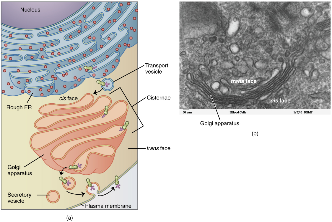
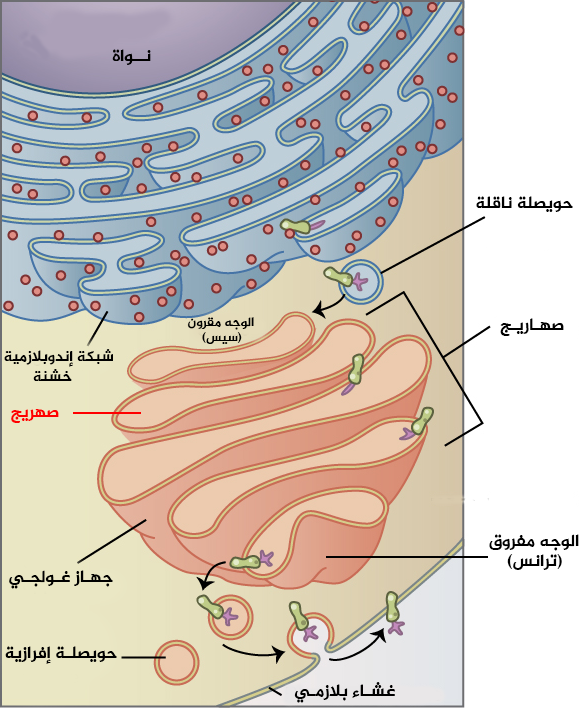
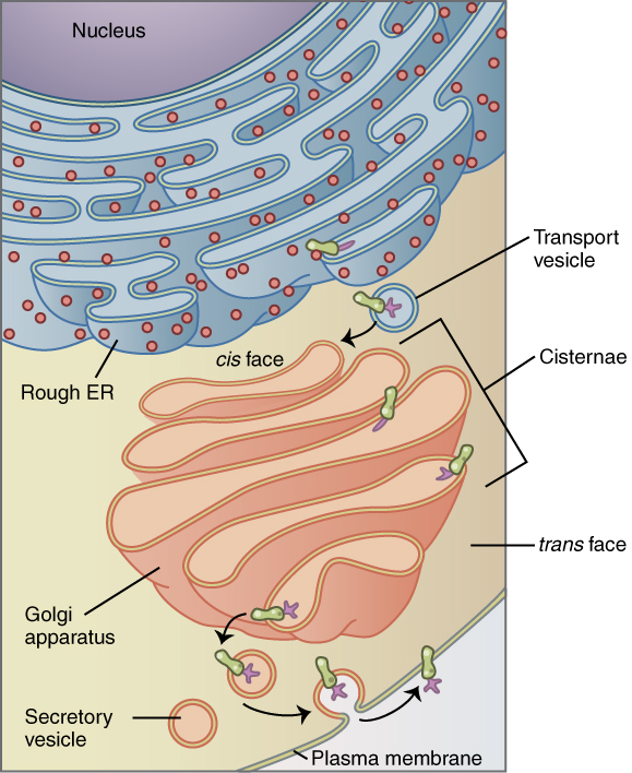
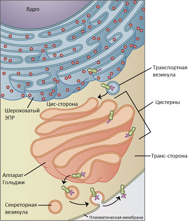
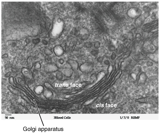
The Golgi apparatus is a major collection and dispatch station of protein products received from the endoplasmic reticulum. Proteins synthesized in the ER are packaged into vesicles, which then fuse with the Golgi apparatus. These cargo proteins are modified and destined for secretion via exocytosis or for use in the cell. The Golgi can be thought of as similar to a post office: it packages and labels items which it then sends to different parts of the cell or to the extracellular space. The Golgi apparatus is also involved in lipid transport and lysosome formation. Its structure and function are intimately linked; individual stacks have different assortments of enzymes, allowing for progressive processing of cargo proteins as they travel from the cisternae to the trans Golgi face. Enzymatic reactions within the Golgi stacks occur exclusively near its membrane surfaces, where enzymes are anchored. Much of the enzymatic processing is post-translational modification of proteins, including glycosylation and phosphorylation. The Golgi apparatus tends to be larger and more numerous in cells that synthesize and secrete large amounts of substances, such as antibody-secreting plasma B cells.
Did You Know?
- The Golgi apparatus was one of the first organelles to be discovered and observed in detail due to its large size and distinctive structure.
- In mammals, a single Golgi apparatus is usually located near the cell nucleus, close to the centrosome.
- The Golgi apparatus adds a mannose-6-phosphate label to proteins destined for lysosomes.
- In some yeasts, such as Saccharomyces cerevisiae, Golgi stacking is not observed.
The Long Road to a Name
In 1898, Italian physician and pathologist Camillo Golgi, while probing the nervous system under his microscope, encountered a striking internal structure he initially labeled the 'internal reticular apparatus.' Its imposing size and distinctive shape meant it was among the first internal structures ever seen clearly under a microscope. Yet the scientific community did not immediately accept the sighting. Critics argued that what Golgi saw was nothing more than an optical artifact produced by his particular observation technique. It took the arrival of modern microscopes during the twentieth century to settle the debate and confirm the structure was genuinely real. The naming history is equally layered. Early references cycled through combinations such as the Golgi–Holmgren apparatus, Golgi–Holmgren ducts, and Golgi–Kopsch apparatus. The phrase 'Golgi apparatus' first entered scientific literature in 1913, and the alternative label 'Golgi complex' did not appear until 1956. The organelle's identity, in other words, was as much a story of institutional consensus as it was of biological discovery.
A Stacked City of Membranes
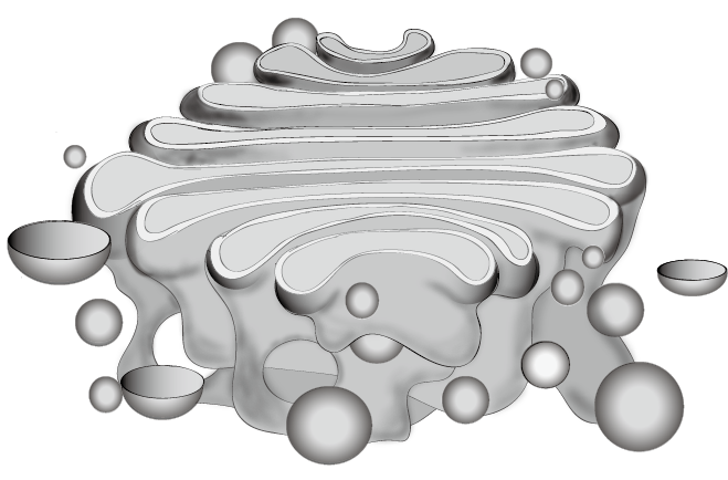
The Golgi apparatus is built from flattened, membrane-enclosed disks called cisternae, sometimes referred to as dictyosomes, which originate as vesicular clusters budding away from the endoplasmic reticulum. In a typical mammalian cell, forty to one hundred of these stacks are present, each holding roughly four to eight cisternae, though certain protists push the count to as many as sixty. Every stack is organized into three functional zones—cis, medial, and trans—collectively forming two larger networks: the cis Golgi network at the entry end and the trans Golgi network at the exit end. The trans network, which may sit adjacent to the stack or float separately, is where proteins are finally sealed into vesicles bound for lysosomes, secretory vesicles, or the plasma membrane. Structural variation across the tree of life is striking. In the yeast Saccharomyces cerevisiae, no stacking is observed at all, while its relative Pichia pastoris does form stacks. Plant Golgi bodies operate as independent units rather than fusing into ribbons. And in cells that must pump out enormous quantities of product, such as antibody-secreting plasma B cells, the Golgi becomes notably larger and more numerous.
The Cell's Post Office
Proteins synthesized in the endoplasmic reticulum are loaded into vesicles that travel to and fuse with the Golgi, where they undergo a series of sequential chemical modifications before being dispatched to their final destinations. The analogy to a postal facility is apt: the Golgi receives raw cargo, applies the correct chemical labels, and routes each parcel to the right address—whether that is a lysosome, a secretory vesicle, or the cell surface. The processing is strictly compartmentalized. In the early cis Golgi network, oligosaccharides on lysosomal proteins are phosphorylated. Moving into the cis cisternae, mannose residues are trimmed away. The medial cisternae handle both further mannose removal and the addition of N-acetylglucosamine. By the time cargo reaches the trans cisternae, galactose and sialic acid are appended. Finally, the trans Golgi network carries out sulfation of tyrosines and carbohydrates. Crucially, all of these enzymatic reactions occur anchored to the membrane surfaces of the cisternae, a design that contrasts sharply with the ER, where many enzymes float freely in the lumen. The Golgi also participates in lipid transport and lysosome formation, and it sits at the crossroads of the secretory, lysosomal, and endocytic pathways.
Where It Sits Depends on Who You Ask
The position of the Golgi apparatus within a cell is far from universal. In mammalian cells, a single Golgi complex typically parks itself near the nucleus, in close proximity to the centrosome, with tubular connections stitching the individual stacks into a continuous ribbon. This architecture is entirely dependent on the microtubule network; when researchers experimentally depolymerize those microtubules, the stacks lose their mutual connections and scatter as isolated units throughout the cytoplasm. Yeast presents a different picture altogether. In Saccharomyces cerevisiae, multiple small Golgi bodies are distributed throughout the cytoplasm rather than consolidated into one structure. Plant cells take yet another approach: their Golgi stacks avoid the centrosomal region entirely, never assemble into ribbon-like structures, and depend on actin cables rather than microtubules to maintain their organization. Despite these differences, one feature is shared across all eukaryotes—every Golgi stack sits adjacent to exit sites of the endoplasmic reticulum, ensuring a constant supply of newly synthesized cargo ready for processing.
Gallery
Frequently Asked Questions
What is the Golgi apparatus?
The Golgi apparatus (also called the Golgi complex or Golgi body) is a membrane-bound organelle found in most eukaryotic cells. It sits within the endomembrane system in the cytoplasm and acts as a central sorting and packaging hub for newly synthesized proteins.
What are the Golgi apparatus's powers/role?
Its core function is to receive proteins from the endoplasmic reticulum, chemically modify them—especially by adding sugar monomers via its resident glycosylation enzymes—and then bundle them into membrane-bound vesicles. Those vesicles are dispatched to their final destinations, whether the cell surface for secretion, a lysosome, or another intracellular compartment.
How does the Golgi apparatus's story end for a given protein?
After a protein has been trimmed, glycosylated, and quality-checked as it traverses the stacked cisternae, the Golgi pinches off a vesicle around it. That vesicle then fuses with its target membrane, delivering the cargo into the secretory, lysosomal, or endocytic pathway it was sorted for.
Why is the Golgi apparatus important?
Without it, cells would lack a central checkpoint where proteins are chemically tailored and correctly addressed before release. It sits at the crossroads of the secretory, lysosomal, and endocytic routes, making it indispensable for cell-to-cell communication, intracellular digestion, and overall membrane homeostasis.
More in Cellular And Molecular Biology 1-24
Elsewhere in the Cellular And Molecular Biology universe
Spotted an error? Know more?
This is a living reference — every entry is fact-audited, and reader corrections feed straight into our audit queue. Suggest an edit · See this site's audit record